Life Cycle of Angiosperms
The Flowering Plants: Phylum Magnoliophyta
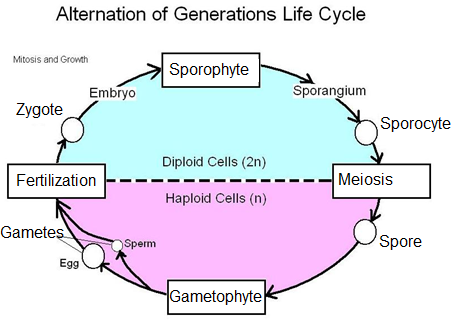
Angiosperms, like all
the higher plants, follow the alternation of generations life cycle.
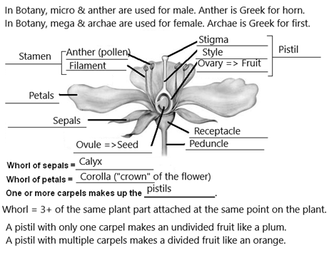
Flower
Structure: We will begin our discussion by reviewing the
structures of the flower.
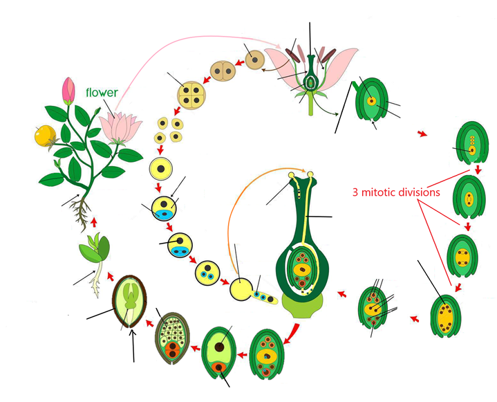
Development of Gametophytes: The diagram below summarizes the development of both the
male and female gametophytes. Recall that gametophytes make gametes. The female
gametophyte only reaches maturity when it produces an egg. The male
gametophyte, likewise, reaches maturity when it produces sperm.
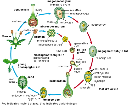
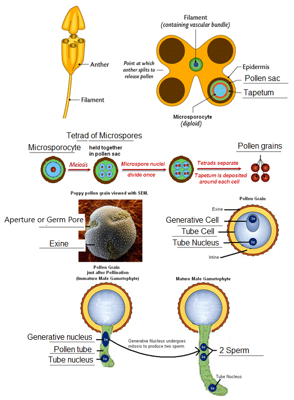
Development of the Male Gametophytes in the
Anther: The anthers are usually made up of two pairs of fused
microsporangia, known as pollen sacs.
Patches of tissue within the microsporangia produce microsporocytes. Microsporocytes undergo meiosis to produce tetrads
of microspores. After meiosis, the
nucleus of each microspore divides once so that the cell has two nuclei. The
tetrads now separate. A two-layered wall develops around each microspore,
resulting in a pollen grain which is
an immature male gametophyte.
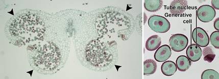
Lily anther and
pollen. Left:
Transverse section of a Lily (Lilium) anther
showing the typical angiosperm arrangement of four pollen sacs (microsporangia)
in two pairs (each pollen sac is indicated by an arrowhead); the sacs contain
2-celled pollen grains. The anther has dehisced (opened) and is ready to
release the pollen. Right: Detail of two-celled pollen grains. The tube cell
will elongate to form the pollen tube, whereas the generative cell will divide
to yield two sperm. Credits: Lilium anther and pollen (CUPAC, copyright 2011 Cornell University Plant Anatomy
Collection, used with permission). Images modified from originals.
Development of the Male Gametophytes in the Anther:
Images modified
from: http://www.ncbi.nlm.nih.gov/bookshelf/br.fcgi?book=dbio&part=A4948 and http://leavingbio.net/TheStructureandFunctionsofFlowers%5B1%5D_files/image005.gif and http://image.tutorvista.com/content/flowering-plants-reproduction/pollen-grain-growth-stages.jpeg
Development
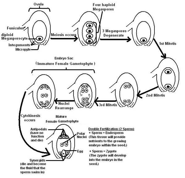
of the Female Gametophyte in the Ovule: Remember
that the ovule will mature to become the seed. Within the ovule of the flower
still developing in its bud, a megasporocyte differentiates from other cells. The megasporocyte undergoes meiosis to produce four megaspores. Only one survives, leaving
the remaining three to degenerate. The nucleus of the remaining megaspore
undergoes three mitotic divisions, yielding a cell that has eight haploid
nuclei. This cell is the immature female gametophyte, referred to as the embryo sac. Four of the nuclei migrate
to the top of this cell as the remaining four nuclei migrate to the bottom. A
single nucleus from each pole of the cell then migrates back to the middle.
These two middle nuclei are the polar
nuclei. Cell walls form around the remaining nuclei at the polls of the
embryo sac. The three cells at the top are the antiopodals.
They serve no purpose and disintegrate. Two of the cells at the bottom are the synergids. The synergids will degenerate and provide the fluid that the
sperm will swim in. The remaining bottom cell grows and becomes the egg. Having produced an egg, this is
now the mature female gametophyte.
During
this process, the outer layers of the ovule differentiate to become the integuments. The integuments will
harden to produce the seed coat, leaving
a small hole known as the micropyle. The micropyle will
allow the pollen tube access to the ovule. Later, this same hole will allow
water to enter the seed, beginning the process of germination.
Development
of the Female Gametophyte in the Ovule:
Pollination: Pollination involves the
transfer of pollen from the anther to the stigma of a flower. Angiosperms are
quite clever in how they deliver the pollen grains to the stigma. Some plants
rely upon gravity, wind, insects, birds, or even mammals to disperse their
pollen to the next plant. Once the pollen becomes stuck on the stigma, it
absorbs fluids and germinates. A pollen
tube bursts from one of the apertures
(also referred to as germ pores) of the pollen grain and begins to grow down
the neck of the pistil, known as the style, then around the ovule to the micropyle.
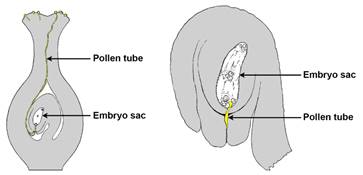
Pollen tube & fertilization. Left: Longitudinal section of a pistil, showing pollen grains on
the stigma. A long pollen tube has grown out of one of the pollen grains and
has made contact with the embryo sac (female gametophyte/megagametophyte)
via the micropyle of the ovule. Right. Detail of an
ovule, showing the pollen tube entering the ovule through the micropyle to penetrate the embryo sac at the time of
fertilization. Credits: Images modified from figures 119 and 231 from Bergen
& Caldwell (1914) Introduction to Botany (no known copyright restrictions).
Fertilization: Fertilization involves the
union of the male and female gamete (the egg and the sperm). It takes at least
24 hours after pollination for the pollen tube to grow all down the style and
reach the micropyle of the embryo sac. In some plant
species, this delay may take as long as one year. Once the pollen tube reaches the
micropyle, it discharges its sperm into the embryo
sac. Angiosperms are unique among plants because they require double fertilization. One sperm
fertilizes the egg, resulting in a zygote.
The other sperm will fertilize the polar nuclei, producing the endosperm. The endosperm becomes a food
tissue utilized in seed development.
The
Seed: After fertilization, the ovule develops into a seed. The
seed contains a young, diploid sporophyte referred to as the embryo. Food required by the embryo to
germinate is stored either in the endosperm or the cotyledons or in both
structures. Seeds often require environmental clues to break their dormancy and
germinate.
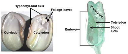
Longitudinal sections of seeds with sporophyte
embryos. Left: Bean (Phaseolus) split lengthwise to show the parts of the
embryo, including the two food-storing cotyledons, the hypocotyl-root axis
(sporophyte embryo axis below the cotyledons), and the first foliage leaves. No
endosperm is apparent. Right: Corn (Zea
mays, a monocot) embryo with one cotyledon and conspicuous endosperm. Credits: Phaseolus seed (Bruce Krichoff,
via flickr, CC
BY 2.0); Zea kernel (Jon Houseman and Matthew
Ford, Wikimedia Commons, CC BY-SA 4.0). Images modified from
originals.
Works
Cited:
“Chapter 23: Seed Plants: Angiosperms.” Introductory
Plant Biology, by James E. Bidlack and Shelley Jansky, McGraw-Hill, 2018, pp. 438–456.
Hermsen, Elizabeth J. “Angiosperm Life Cycle.” Digital
Atlas of Ancient Life, Digital Atlas of Ancient Life Paleontological
Research Institution, 9 Aug. 2019,
www.digitalatlasofancientlife.org/learn/embryophytes/angiosperms/angiosperm_life_cycle/.
Life Cycle of Angiosperms